[ad_1]
New study reveals that PFA ependymoma brain tumors exhibit unique 3D genomic features that could be exploited for therapeutic purposes
Montreal, July 10, 2024 – Group A posterior fossa ependymoma (PFA) is a rare, treatment-resistant pediatric tumor of the central nervous system that originates in the brain and spinal cord. It has the highest recurrence rate and poorest prognosis of all childhood cancers, due to the lack of effective treatment.
Hope is on the horizon now that an international research team led by scientists from Baylor College of Medicine in Texas, USA, and the Research Institute of the McGill University Health Centre (RI-MUHC) in Montreal, Canada, have identified unique three-dimensional features called TULIPs in the PFA ependymoma genome that could potentially be targeted in the development of more effective therapies. The findings are published in Cell.
“PFA ependymomas are deadly. One reason for the lack of progress in developing effective treatments for these tumors is that the majority of PFAs do not have clear genetic mutations that drive tumor growth. Without a clear genetic target against which we could design specific therapies, we studied another aspect of the tumor: how DNA is packaged inside the cell nucleus,” said lead and senior author, Marco GalloAssociate Professor of Pediatrics, Hematology-Oncology at Baylor and Texas Children’s Hospital.
“Our work was motivated by a simple observation: plantar aortic ependymomas are generally diagnosed in very young children and there is no effective treatment. Radiation therapy, the only treatment currently available, is not effective and leads to serious developmental and cognitive problems. This is a reality that we hope to change,” says the Canada Research Chair in Pediatric Oncology Dr. Nada Jabadoco-senior author of the study, senior scientist in the Child Health and Human Development program at the RI-MUHC and pediatric hematologist-oncologist at the Montreal Children’s Hospital of the MUHC.
Discovering the 3D configuration of tumor cell genomes
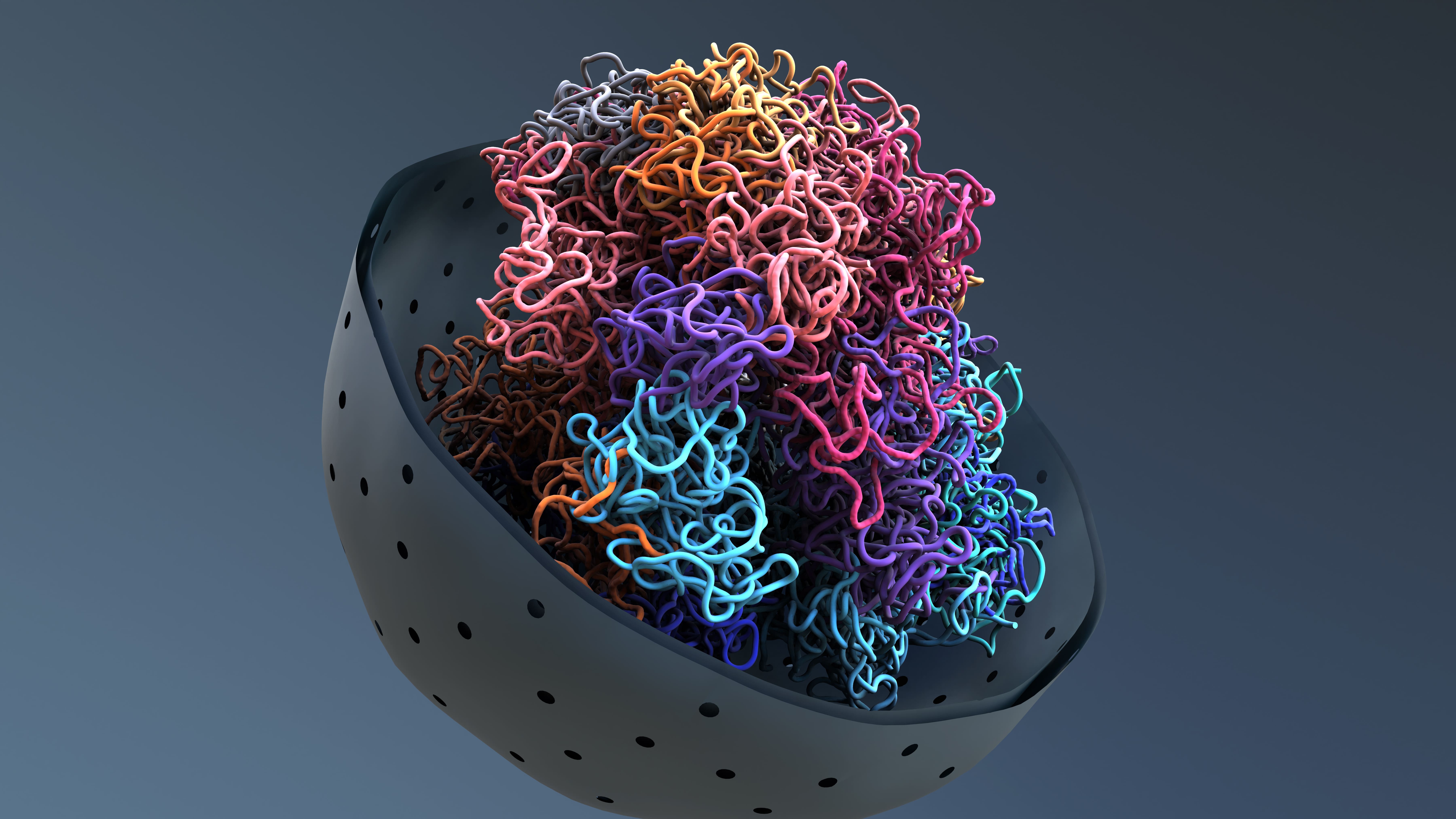 Every cell in the body has about 2 meters of linear DNA that is stored in its nucleus so that the cell can easily access the genes it uses most often and set aside the ones it uses less often. This would be like organizing the closet with the most frequently used clothes in the front and the rarely worn ones in the back. To fit into the tiny nucleus, the long DNA molecules are bent, twisted, and looped, resulting in specific 3D conformations, some tighter, some more relaxed, that can ultimately help the cell express the genes it needs to do its job.
In this study, the researchers took a close look at what we might call the “geography” of the PFA ependymoma cell genome (the complete set of DNA instructions found within the cell).
“We studied the unique ways in which PFA cells organize their DNA in 3D, orchestrating strong interactions between regions of the genome that are normally far apart. We discovered specific regions that are not present in other types of pediatric brain cancer and that reappear at predictable genomic locations. We called them TULIPs, for Type B Ultra-Long Interactions in PFAs,” explains Dr Michael D Taylorco-senior author of the study and professor of pediatrics, hematology-oncology and neurosurgery at Baylor and Texas Children’s. He also holds the Cyvia and Melvyn Wolff Chair in Pediatric Neuro-Oncology at Texas Children’s Cancer and Hematology Center.
The researchers used Hi-C technology to profile the 3D architectures of the entire genomes of PFA tumors and compared them to those of a large cohort of samples from different tumor types and non-malignant tissues. In the process, TULIPs emerged as specific regions of DNA that were tightly packed and therefore difficult to access, a sign that the cell may not use genes in that region often.

“TULIPs also tend to interact with each other over very long distances. One TULIP can be at one end of a chromosome and another TULIP at the other end of the same chromosome, and they manage to interact with each other with surprising strength,” says Professor Gallo. “TULIPs on different chromosomes can also converge and interact strongly with each other. We also found that regions outside of TULIPs appear more relaxed overall. This is important because TULIPs are linked to the function of the cell.”
A potentially exploitable chemical label
According to the study results, TULIPs carry a methyl group on histone H3K9, a DNA-associated protein that can act as a chemical marker. Indeed, when the research team inhibited H3K9 tagging in cultures derived from AFP patients, they found weaker interactions between TULIPs and impaired AFP cell survival. These observations suggest that TULIP interactions are important for AFP cell viability, opening up new potential targets for treatment.
“We believe that TULIPs are ephemeral structures present at an early stage of cancer development in progenitor cells, that is, cells that descend from stem cells and precede the creation of mature cells, very early in life. However, further research is needed to understand the mechanism by which TULIPs arise and govern the behavior of cancer cells,” says Dr. Jabado, who is also a professor in the Department of Pediatrics at McGill University. “By further studying this mechanism, we may be able to explore treatment strategies to eliminate them in order to promote tumor elimination.”
About the study
The study TULIPs decorate the three-dimensional genome of PFA ependymoma was conducted by Michael J Johnston, John JY Lee, Bo Hu, Ana Nikolic, Elham Hasheminasabgorji, Audrey Baguette, Seungil Paik, Haifen Chen, Sachin Kumar, Carol CL Chen, Selin Jessa, Polina Balin, Vernon Fong, Melissa Zwaig, Antony MichealRaj, Xun Chen, Yanlin Zhang, Srinidhi Varadharajan, Pierre Billon, Nikoleta Juretic, Craig Daniels, Amulya Nageswara Rao, Caterina Giannini, Eric M Thompson, Miklos Garami, Peter Hauser, Timea Pocza, Young Shin Ra, Byung-Kyu Cho, Seung-Ki Kim, Kyu-Chang Wang, Ji Yeoun Lee, Wieslawa Grajkowska, Marta Perek-Polnik, Sameer Agnihotri, Stephen Mack, Benjamin Ellezam, Alex Weil, Jeremy Rich, Guillaume Bourque, Jennifer A Chan, V Wee Yong, Mathieu Lupien, Jiannis Ragoussis, Claudia Kleinman, Jacek Majewski, Mathieu Blanchette, Nada Jabado, Michael D Taylor and Marco Gallo.
DO I: https://doi.org/10.1016/j.cell.2024.06.023
This work was supported by a Large-Scale Applied Research Project Grant from Génome Québec, Genome Canada, the Government of Canada, and the Ministère de l’Économie et de l’Innovation du Québec, with support from the Ontario Research Fund through funding provided by the Government of Ontario. Additional support was provided by Brain Canada through the Canada Brain Research Fund, Health Canada, and the Azrieli Foundation through an Azrieli Future Leader in Canadian Brain Research Grant, Canadian Institutes of Health Research (CIHR) Project Grants (PJT-156278 and PJT-173475), a CIHR Postdoctoral Fellowship, and a Canada Research Chair.
About the IR-CUSM
The Research Institute of the McGill University Health Centre (RI-MUHC) is a world-renowned biomedical and health research centre. The Institute, which is affiliated with the Faculty of Medicine of McGill University, is the research arm of the McGill University Health Centre (MUHC) – an academic health centre located in Montreal, Canada, with a mandate to focus on complex care within its community. The RI-MUHC supports more than 720 researchers and nearly 1,400 research trainees who are engaged in a broad range of basic, clinical and health outcomes research at the Glen and Montreal General Hospital sites of the MUHC. Its research facilities provide a dynamic, multidisciplinary environment that fosters collaboration and leverages discoveries to improve the health of patients throughout their lives. The RI-MUHC is funded in part by the Fonds de recherche du Québec – Santé (FRQS). www.rimuhc.ca
/Public dissemination. This content from the original organization/authors may be of a timely nature and edited for clarity, style, and length. Mirage.News takes no institutional position or bias, and all views, positions, and conclusions expressed herein are solely those of the author(s). See the full story here.